Cutting edge of mechanical imaging | Tsinghua team achieves new breakthrough in synchronous non-invasive measurement of multiple mechanical parameters of arteries!
Cardiovascular disease is one of the major health burdens worldwide.Biomechanical indicators of arteries, including intramural stress, axial/circumferential stiffness and local blood pressure, are not only the physiological basis for maintaining normal hemodynamics, but also important mechanical clues for the occurrence and progress of hypertension, atherosclerosis, heart failure, stroke and other diseases. These indicators are highly dynamically coupled in time and space and exhibit significant anisotropy,However, existing in vivo measurement methods are mostly limited to a single indicator or rely on multiple measurements at different times/locations and population statistical assumptions, making it difficult to achieve local, synchronous, and individualized mechanical characterization.
PART 01
New research, new progress
New Research & New Developments
In August 2025, Professor Cao Yanping's research group from the Institute of Biomechanics and Medical Engineering at the School of Aeronautics and Astronautics, Tsinghua University, in collaboration with Li Guoyang's research group at Peking University and Wang Xinyu's team at the Third Hospital of Peking University, published a groundbreaking study in Science Advances.This study proposes and validates a "same position and same phase" guided wave elastography method based on programmed acoustic radiation force and ultrafast ultrasound imaging, which achieves in vivo, non-invasive, and synchronous measurement of arterial circumferential and axial guided waves, and uses this to invert local blood pressure, bidirectional wall stress, and anisotropic stiffness. This method provides a new mechanical imaging tool for studying arterial mechanical coupling, physiological pathological changes, and precise clinical evaluation.
Welcome to read the original text for more technical details and data support: https://doi.org/10.1126/sciadv.adv5660
This study utilized programmed acoustic radiation force to non invasively excite elastic guided waves propagating along both axial and circumferential directions in the blood vessel wall using serialized focused pulses (Figure 1A-B). And real-time capture of the propagation process of bidirectional guided waves in the same longitudinal section is achieved through an ultra fast ultrasound imaging system (frame rate ≥ 8000 Hz) (Figure 1C-E), obtaining the spatial temporal velocity field of bidirectional guided waves. Based on the wavefield information, the team constructed a guided wave dynamics model that integrates vascular viscoelasticity, finite deformation, and actual boundary conditions, and developed an intelligent inversion algorithm. Thus achieving:
- Continuous non-invasive estimation of blood pressure:The study found a significant correlation between the characteristics of circumferential guided waves and local blood pressure (Figure 2A-B). In experiments with different positions (such as neck extension), the sensitivity of circumferential waveguide velocity to posture is much lower than that of axial wave velocity, showing better robustness (Figure 2C). Compared with the percutaneous flattening method, this method has good comparability in the volunteer population (Figure 2D-E), and this research method does not require compression of blood vessels, significantly improving comfort and applicability.
- Synchronous inversion of stress and stiffness:By dynamically changing the wave properties such as the dispersion characteristics of axial and circumferential guided waves (Figure 3A, 3C, 3G), the bidirectional stress (Figure 3D-E, 3H-I) and stiffness (Figure 3F, 3J) of the vascular wall during diastole and systole can be inverted. Under the posture of neck extension, the axial stress significantly increases (about 13 kPa), while the circumferential stress changes less, highlighting the sensitivity and accuracy of this technique to changes in mechanical states (Figure 3D). The population data shows that the circumferential stress of the carotid artery is generally higher than the axial stress, and the circumferential stiffness is significantly greater than the axial stiffness, indicating that the mechanical properties of the arterial wall have strong anisotropy (Figure 3E – F, 3H – J).
- Measurement during cardiac cycle and pathological sensitivity:This study only requires about 4 milliseconds for a single data acquisition, supporting real-time tracking of changes in mechanical parameters within one or more cardiac cycles. The in vivo experimental results showed that there were significant differences in the mechanical characteristics of volunteers in the youth group, middle-aged and elderly normal blood pressure group, and middle-aged and elderly hypertension group, especially in the hypertension group where the circumferential stiffness and stress increased significantly, indicating the high sensitivity of this method in evaluating the vascular health status of this population (Figure 3G-J).
PART 02
Research Results Diagram
Research Findings Diagram
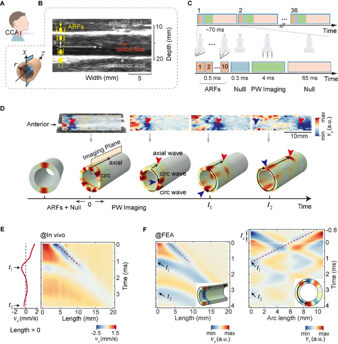
Figure 1: Schematic diagram of bidirectional guided wave excitation and measurement experiment and in vivo data
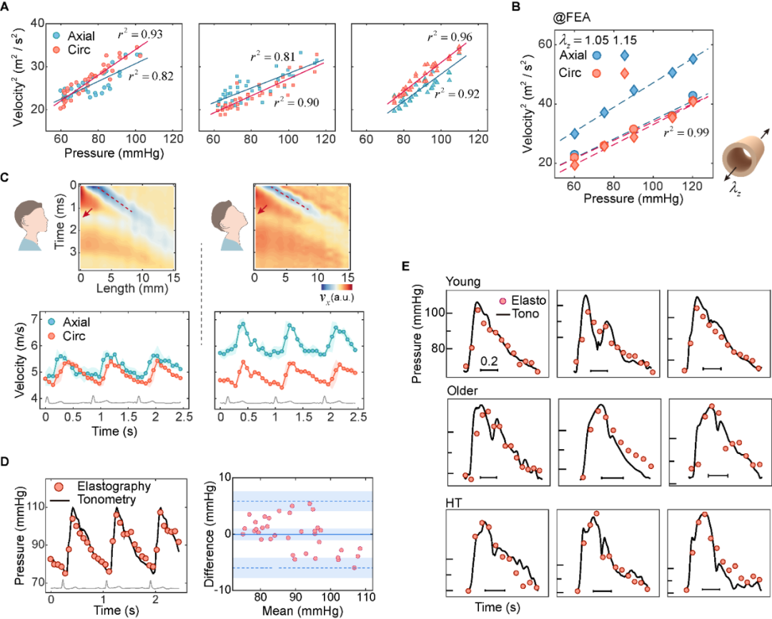
Figure 2: Comparison of blood pressure measurement results with flattening method shows excellent consistency
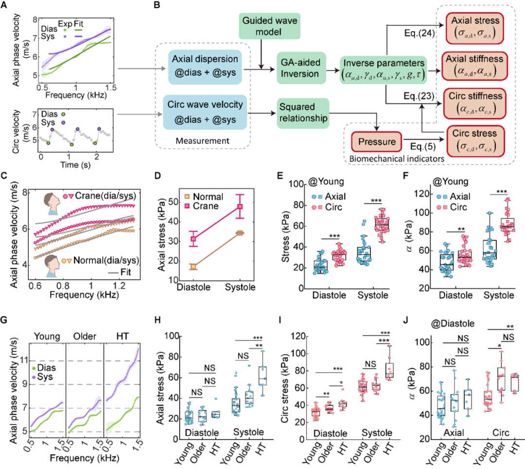
Figure 3: Characterization results of arterial stress and stiffness in various postures and populations
PART 03
Conclusions and Prospects
Conclusion and Outlook
This study achieved a breakthrough in in vivo, synchronous, and non-invasive measurement of multiple mechanical parameters of arteries, solving the key bottlenecks of spatiotemporal inconsistency and dependence on prior parameters in previous technologies.This method not only has the potential to reveal the mechanisms of vascular pathological aging and remodeling, but also provides a new mechanical approach for early screening, dynamic monitoring, and personalized evaluation of cardiovascular diseases. In the future, this technological framework can also be extended to fields such as cardiac mechanical imaging, artificial blood vessel evaluation, and flexible device mechanical monitoring, with broad clinical application prospects.
PART 04
Collaboration Team and Author Introduction
Research Team & Author Introduction
This study was led by Professor Yanping Cao's research group at the Institute of Biomechanics and Medical Engineering, School of Aerospace Science and Technology, Tsinghua University.Cao Yanping, a professor at the Institute of Biomechanics and Medical Engineering of the School of Aerospace Science at Tsinghua University, Li Guoyang, an assistant professor at the School of Mechanics and Engineering Sciences at Peking University, and Wang Xinyu, chief physician of the Department of Cardiology at Peking University Third Hospital, are the co corresponding authors of the paper. The first author of the paper is Dr. Jiang Yuxuan from Cao Yanping's research group (currently a postdoctoral fellow at Harvard Medical School). Other authors include Hu Keshuai, Ma Shiyu, Dr. Zheng Yang (currently serving as the Chief Technology Officer of Xijian Technology), Master Jiang Mingwei, and Zhang Zhaoyi, members of Cao Yanping's research group.This research is supported by projects such as the National Natural Science Foundation of China, and the core results have been applied for national invention patent protection.
This study is part of the research content of Dr. Jiang Yuxuan, the first author of the paper. Dr. Jiang Yuxuan focused on the mechanical imaging theory and imaging methods of the cardiovascular system during his doctoral studies, and developed a series of in vivo imaging methods for arteries, veins, and the heart. As the first or co first author, he published 5 academic papers and granted 4 invention patents in authoritative journals such as Sci. Adv., IEEE Trans. Med. Imaging, and Int. J. Eng. Sci,One of them has been transferred to Xijian TechnologyDr. Jiang Yuxuan is currently actively promoting research on cardiovascular imaging technology, aiming to achieve higher resolution vascular mechanical characterization and explore its application prospects in clinical diagnosis.